

Why This Study Matters Right Now

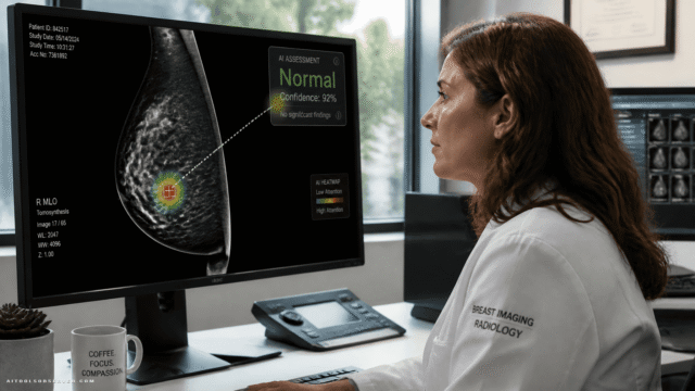
Lung cancer is one of the deadliest cancers globally. Chest radiography is typically the first line of investigation when symptoms appear, which makes accurate AI-assisted interpretation a genuine life-or-death question.
The UK’s National Optimal Lung Cancer Pathway mandates that chest X-rays from primary care be reported within 24 hours. But a 29% shortfall in radiologist numbers is making that target increasingly difficult to hit.
That’s exactly why AI tools have entered the conversation — to prioritise urgent scans, support same-day CT referral decisions, and catch subtle early-stage tumours that human readers might miss under pressure. In 2023, the UK government committed £21 million across 12 imaging networks in England to accelerate AI deployment, including lung cancer detection tools.
The problem? NICE previously concluded there wasn’t enough evidence to support routine clinical adoption. This study is the most direct test of that concern yet.
How the Study Was Designed
Researchers conducted a retrospective analysis of 5,235 consecutive adult patients who had chest radiographs taken between July 2020 and February 2021 at a single UK centre.
Key patient demographics:
- Median age: 60 years
- Gender split: 53.4% women
- Ethnicity: 79.4% White
- Confirmed lung cancer prevalence: 1.4% (with a visible tumour, verified by multidisciplinary team diagnosis as the reference standard)
Every radiograph was independently analysed by all seven AI systems — no human radiologist input, no cross-contamination between tools. This is as clean a head-to-head comparison as you’re likely to see in clinical AI research.
The Numbers: A Wide and Worrying Performance Gap

This is where the study gets uncomfortable for vendors and procurement teams alike.
Diagnostic Accuracy at a Glance

| Metric | Lowest Performer | Highest Performer |
|---|---|---|
| AUC (ROC curve) | 0.80 | 0.94 |
| Sensitivity | 20.8% | 77.8% |
| Specificity | 58.9% | 98.4% |
| Positive Predictive Value | 1.5% | 28.4% |
| Additional False Positives | 10 cases | 2,039 cases |
Let that sink in. The worst-performing tool in terms of sensitivity caught fewer than 1 in 4 lung cancers. The worst in terms of false positives generated over 2,000 incorrect flags — in a dataset of just over 5,000 patients.
Compared to Radiologists
Three of the seven AI tools identified more tumours than radiologists did. That sounds like a win — until you factor in the false-positive burden those tools carried.
Four tools detected fewer cancers than radiologists, which raises an obvious question: what exactly are hospitals paying for?
The False Positive Problem Is Not a Minor Detail

False positives in cancer screening aren’t just a statistical inconvenience. Each one triggers follow-up imaging, patient anxiety, clinical time, and downstream costs.
A tool generating 2,039 additional false positives in a 5,235-patient cohort would, at scale across an NHS imaging network, create an enormous secondary workload. That directly undermines the original goal of using AI to reduce pressure on radiology departments.
This is the core tension in deploying sensitivity-optimised tools without accounting for specificity. Higher sensitivity often means catching more true cancers — but it also means flagging more healthy patients as suspicious.
The right balance depends entirely on the clinical context, the downstream pathway, and the capacity of the system to handle follow-up investigations.
These Tools Are Not Interchangeable

One of the study’s most significant findings is often buried in the technical language: agreement between the seven AI systems was minimal.
Different platforms frequently flagged entirely different patients as suspicious for lung cancer. This isn’t a minor calibration difference — it means two hospitals using different AI tools on the same patient population would likely produce substantially different referral lists.
For procurement teams and clinical leads, this has a direct implication: you cannot assume that any CE-marked or FDA-cleared lung cancer AI tool will perform comparably to another. Regulatory clearance is not a performance guarantee.
What This Means for NHS AI Adoption

The NHS is actively expanding investment in imaging AI. But this study reinforces what NICE flagged earlier — the evidence base for routine clinical use remains thin, and the variability between commercial products is real and consequential.
The researchers explicitly called for further studies to assess how these tools influence:
- Radiologist performance — do they help or anchor human readers to incorrect outputs?
- Patient outcomes — does AI-assisted detection actually improve survival rates?
- Service delivery — does the net effect reduce or increase workload?
These are not abstract academic questions. They are the questions that should be answered before any health system commits to large-scale AI adoption.
How to Use This Study When Evaluating Radiology AI Tools

If you’re involved in AI procurement, clinical governance, or health tech strategy, here’s what this research tells you practically:
1. Demand independent validation data, not vendor benchmarks.
The performance gap between tools in this study was enormous. Internal vendor testing on curated datasets will almost always look better than real-world performance on your patient population.
2. Evaluate sensitivity and specificity together — never in isolation.
A tool with 77.8% sensitivity sounds impressive. A tool with 77.8% sensitivity and 2,039 false positives per 5,000 patients is a workflow problem.
3. Test on your own data before committing.
This study used a single UK centre’s patient population. Demographics, equipment, and imaging protocols vary. Performance will vary too.
4. Don’t assume regulatory clearance equals clinical equivalence.
All seven tools in this study were commercially available. Their performance ranged from genuinely useful to potentially harmful in a clinical setting.
5. Plan for the false positive pathway before deployment.
If your AI tool flags 10% of scans as suspicious, what happens next? Who reviews them? How fast? At what cost? These questions need answers before go-live.
The Bigger Picture for AI Tool Buyers

This study is a case study in why independent, real-world comparisons matter — and why the AI tools market needs more of them.
Marketing materials for radiology AI tools routinely lead with AUC scores and sensitivity figures from internal validation studies. What this research shows is that those numbers can look very different when seven tools are tested side by side on the same 5,235 patients.
The AUC range of 0.80 to 0.94 might seem narrow in isolation. But in clinical practice, the difference between a tool that catches 20.8% of lung cancers and one that catches 77.8% is the difference between a useful clinical aid and a dangerous false sense of security.
Final Takeaway
The AI radiology market is maturing fast — but this study is a clear signal that maturity in marketing hasn’t yet translated to consistency in performance.
For NHS trusts, imaging networks, and any health system evaluating lung cancer AI tools: the burden of proof sits with the vendor, not the buyer. Demand independent validation. Test on your own population. And never treat two tools designed for the same task as clinically equivalent until the data says otherwise.
The tools that perform best in controlled studies aren’t always the ones with the biggest marketing budgets. Observe carefully. Choose smarter.


Comments (0) No comments yet
Want to join this discussion? Login or Register.
No comments yet. Be the first to share your thoughts!