
The Problem With Conventional Spatial Profiling

Understanding how genes are expressed across different regions of a tumor is not a trivial task. Tumors are biologically heterogeneous: gene activity in one section can differ substantially from another, and those differences carry real clinical weight — influencing prognosis, treatment response, and resistance mechanisms.
Traditional spatial gene expression profiling captures this complexity, but at a steep price. The process typically takes several weeks and costs thousands of dollars per sample. That combination of time and expense has kept cohort sizes small and clinical adoption essentially out of reach.
Before Path2Space existed, the largest dataset available for studying spatial tumor organization contained roughly 30 patients. That is not a foundation on which scalable precision oncology can be built.
What Path2Space Actually Does

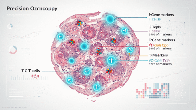
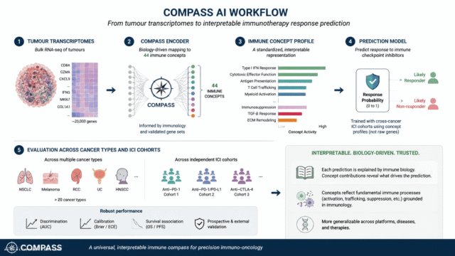
Path2Space works from digital images of standard biopsy slides — thin tissue sections already routinely prepared in clinical settings. From those images alone, the model predicts spatial gene expression across the tumor area, estimating activity at multiple distinct points rather than producing a single averaged readout.
The key output is the prediction of nearly 5,000 genes per sample, mapped spatially across the tumor. Validation against three independent patient datasets showed that predictions aligned well with directly measured gene expression — a result that establishes the model’s reliability beyond its training cohort.
The process takes minutes, not weeks. The cost is a fraction of conventional methods.
Why Spatial Resolution Matters

The distinction between bulk gene expression and spatial gene expression is not merely technical — it is clinically meaningful. A gene expressed uniformly across a tumor tells a different story than one expressed only in isolated pockets. Those spatial patterns can indicate how a tumor is likely to behave, how it may respond to therapy, and which patients face elevated risk of poor outcomes.
Path2Space is explicitly designed to surface these patterns. The research team identified specific spatial signatures of gene activity that correlated with treatment response — findings that would have been statistically invisible in small cohorts or bulk expression data.
This is where the tool’s value compounds: it does not just accelerate existing workflows. It opens analytical territory that was previously inaccessible.
Scale as a Scientific Multiplier

The shift from 30-patient cohorts to thousands is not incremental — it is transformative for the statistical power available to researchers. Biomarker discovery, in particular, depends on large, well-characterized datasets to distinguish signal from noise.
With Path2Space, researchers can now apply spatial analysis retrospectively to existing slide archives, dramatically expanding the data available for study without requiring new tissue collection or expensive re-profiling. The tool effectively unlocks the latent value of pathology archives that institutions have accumulated over decades.
This scalability also positions Path2Space as infrastructure for future research, not just a one-time analytical improvement.
Expanding Scope: Beyond Breast Cancer

The initial validation focused on breast cancer, where both biopsy slides and outcome data were available at sufficient scale. However, the architecture is not disease-specific. Path2Space can be retrained on data from other cancer types, and the Cedars-Sinai team is already finalizing a study applying the model to head and neck cancer.
One current technical limitation is resolution: the model currently analyzes groups of 10 to 20 cells together rather than individual cells. The team is actively working to improve granularity, with single-cell resolution as the stated goal. That improvement would further sharpen the clinical utility of spatial predictions.
The Clinical Pathway Ahead
Path2Space has not yet entered clinical trials. The research team is clear-eyed about the validation work that remains before the tool can influence treatment decisions directly. Eytan Ruppin, MD, PhD, the study’s senior author and deputy director of the Translational Research Institute at Cedars-Sinai, has framed clinical trial validation as the next essential step.
The path from research tool to clinical instrument is neither short nor guaranteed. But the foundational requirements — predictive accuracy, scalability, cost efficiency, and applicability to standard clinical materials — are already in place.
If clinical validation confirms the tool’s performance, Path2Space could make spatial tumor profiling a routine component of cancer workup rather than a specialized research procedure available only at well-funded institutions.
What This Means for AI in Precision Oncology

Path2Space is a precise example of what productive AI integration in medicine looks like: a model trained on a specific, high-value task, validated rigorously against independent datasets, and designed to reduce a real bottleneck rather than automate a peripheral function.
The broader implication is structural. As tools like Path2Space mature, the cost and time barriers that have historically limited spatial biology will erode. Researchers will gain access to larger, richer datasets. Clinicians will gain access to more granular tumor characterization. Patients — particularly those outside major academic medical centers — may gain access to precision oncology approaches that currently require resources most institutions do not have.
The gap between what spatial transcriptomics can reveal and what clinical practice can act on has been a persistent constraint. Path2Space is a credible step toward closing it.


Comments (0) No comments yet
Want to join this discussion? Login or Register.
No comments yet. Be the first to share your thoughts!