


What the AI Actually Does
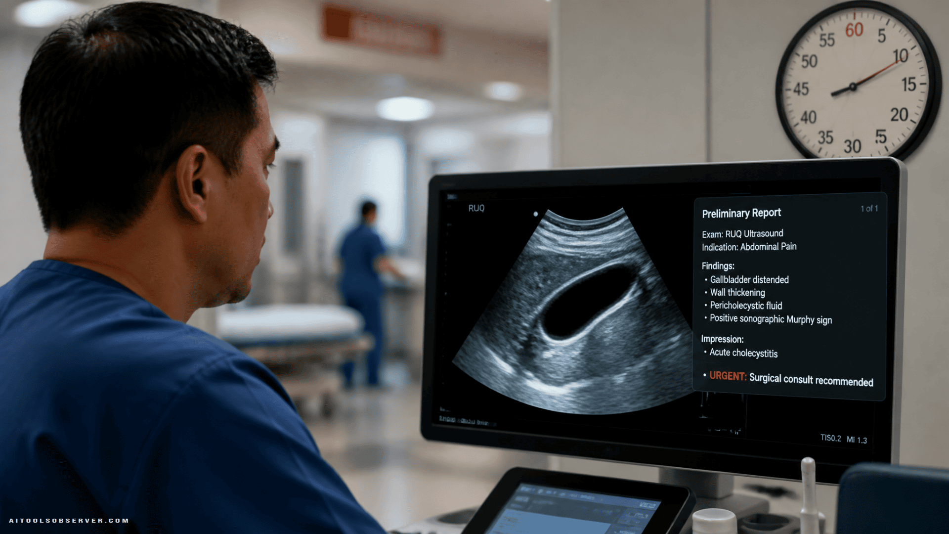
RUQ ultrasounds visualize the liver, gallbladder, right kidney, and part of the pancreas. They’re ordered when patients present with upper abdominal pain or abnormal lab results like elevated liver enzymes or bilirubin. Interpreting them accurately takes skill, and that skill varies widely across practitioners.
The VLM the researchers built combines visual ultrasound data with written clinical information—lab results, patient context—to produce a complete diagnostic picture. Specifically, the model is designed to:
- Scan ultrasounds for 16 common diagnoses
- Estimate each patient’s prognosis
- Auto-generate preliminary radiology reports
- Rapidly flag urgent cases like acute cholecystitis, which requires emergency surgery
The team tested the model on roughly 11,000 RUQ ultrasounds sourced from Stanford University, the University of Colorado, and Johns Hopkins University. They then manually reviewed 100 AI-generated reports for accuracy.
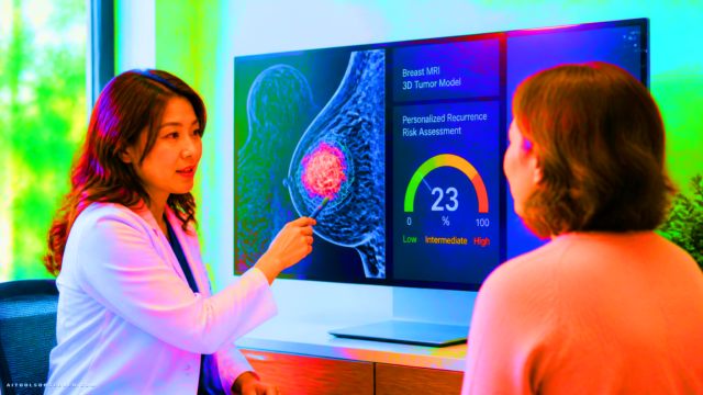
This sits within a broader shift in medical imaging workflows.
The Results Worth Paying Attention To
In that reviewed sample, the VLM identified correct diagnoses 99.6% of the time. That’s a strong signal, though it’s worth noting this was a controlled evaluation, not a live emergency department deployment.
The more telling finding came from a blinded expert review. A panel of specialists was asked to distinguish between reports written by the AI and reports written by radiologists. They could identify unedited AI reports. But when radiologists reviewed and lightly edited the AI-generated reports, the experts could no longer reliably tell them apart from fully human-written ones.
That’s a meaningful result. It suggests the AI isn’t producing rough drafts that need heavy rework—it’s producing outputs that, with a quick review pass, meet professional standards.
Why Radiology Specifically Needs This
The workforce pressure in radiology is real and measurable. An aging population is driving higher demand for medical imaging. At the same time, radiologists are leaving the field at roughly twice the rate they were in 2014. Reimbursement from Medicare has also declined, compressing the economics further.
The result is a specialty where practitioners are being asked to read more images, write more reports, and do it faster—with fewer colleagues to share the load.
If an AI tool can handle the first draft of a report, radiologists can focus their attention on review, edge cases, and clinical judgment. That’s a workflow shift, not a replacement.
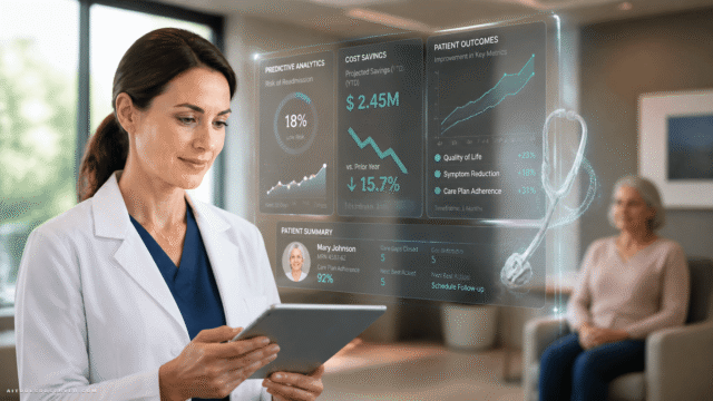
This kind of support is especially relevant to radiology workflow.
The Collaboration Model, Not the Replacement Model
Senior author Harrison Bai, MD, MS, is direct about the intent: this is not an AI radiologist. The VLM is designed to work alongside trained radiologists, not instead of them. It generates the preliminary report; the radiologist reads, edits, and signs off.
That framing matters for adoption. Radiologists are more likely to use a tool that saves them time than one that feels like it’s competing with their role. The researchers designed the system with that psychology in mind.
The model is also positioned as a “clinically integrated assistant”—one that can pull together different data types (imaging plus lab values) and support decision-making without overriding clinical expertise.
What Still Needs to Happen Before Widespread Use
The researchers are clear that this work is promising but not finished. A few things need to happen before this technology moves into routine emergency care:
- Real-world validation: Performance in a controlled study doesn’t automatically translate to a busy, chaotic emergency department.
- Physician-AI collaboration studies: How does working with this tool actually affect diagnostic accuracy and efficiency in practice?
- Patient outcome tracking: Does faster reporting lead to better patient outcomes, or does it introduce new failure modes?
These aren’t reasons to dismiss the research—they’re the standard bar for any clinical tool moving from lab to practice.
The Practical Takeaway
This research points to a specific, credible use case for AI in medicine: not replacing clinical judgment, but reducing the time it takes to get from image to insight. For emergency diagnoses where minutes matter—acute gallbladder inflammation being the clearest example—a tool that generates an accurate preliminary report in seconds has real clinical value.
The vision-language model approach is also worth watching more broadly. Combining image data with structured clinical text is a pattern that could extend well beyond abdominal ultrasounds into other imaging modalities where report generation is a bottleneck.
For anyone tracking AI tools in healthcare, this is a useful benchmark: what good AI-assisted radiology looks like isn’t a system that works alone—it’s one that makes the radiologist faster without making them redundant.








Comments (0) No comments yet
Want to join this discussion? Login or Register.
No comments yet. Be the first to share your thoughts!